
Research Focus
3D-Printed Cardiovascular Implants and Cardiac Patches

We develop polymer-based cardiovascular implants using advanced additive manufacturing and 3D-printing technologies. A major focus is the fabrication of cardiac patches for myocardial repair. Their architecture and viscoelastic properties are tailored to reproduce the complex, nonlinear, and anisotropic mechanical behavior of native cardiac tissue. These patches are designed to mechanically stabilize infarcted regions, limit adverse cardiac remodeling, and support tissue repair.
Biofunctionalization, Coating, and Encapsulation

We use coating and encapsulation strategies to equip cardiovascular implants with specific biological functions. Extracellular matrix components are incorporated to create cell-instructive surfaces that support cell adhesion, proliferation, maturation, and tissue integration. Bioactive molecules, including nitric oxide donors, are integrated or encapsulated for controlled and localized release. These approaches aim to promote endothelialisation and reduce adverse vascular responses, such as intimal hyperplasia.
Nano- and Micropatterned Surfaces for Blood-Contacting Implants
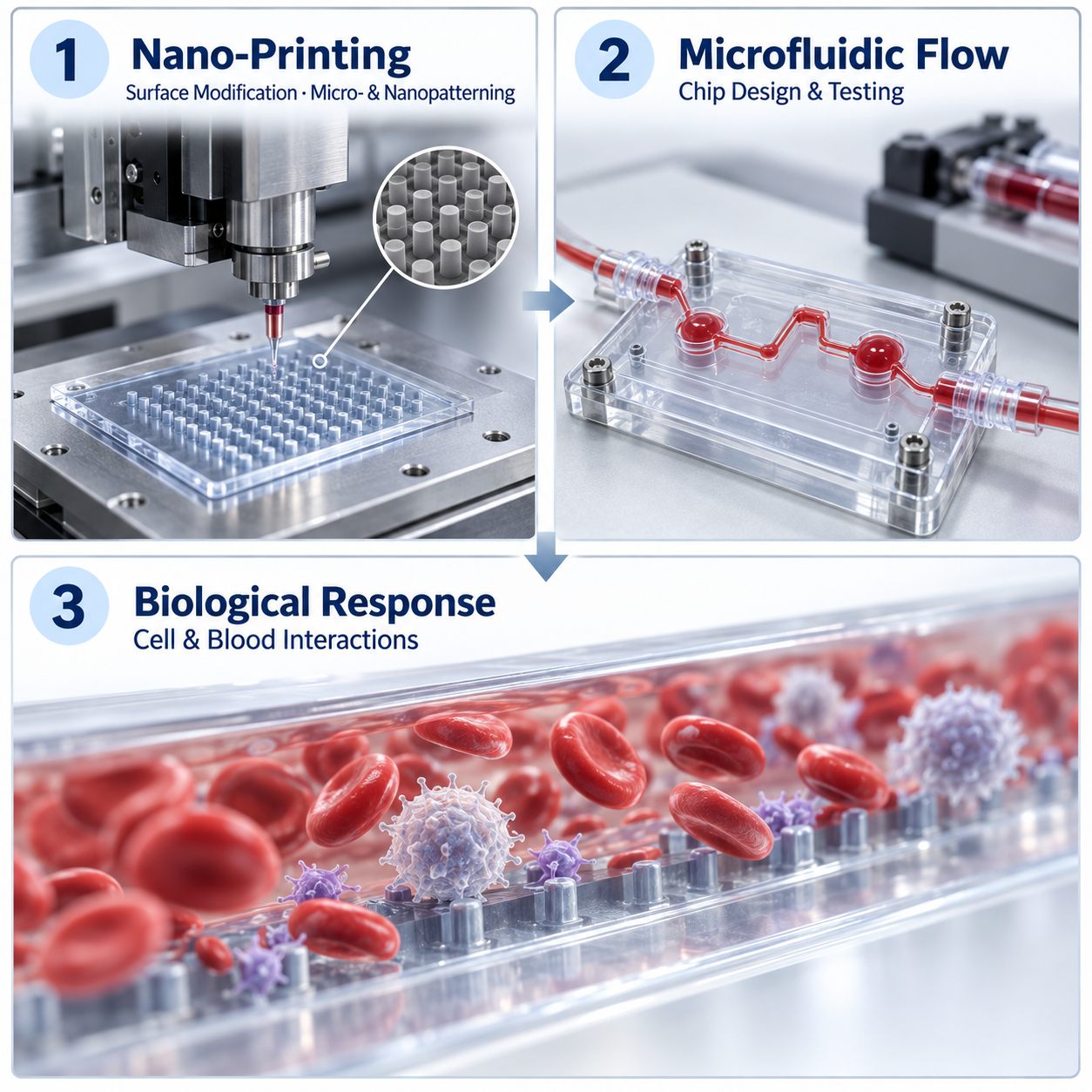
In collaboration with Univ.-Prof. Francesco Moscato and with full funding from the Ludwig Boltzmann Institute for Cardiovascular Research (LBI), nano- and micropatterned implant surfaces are fabricated using high-resolution nano-printing. Dynamic flow models are used to assess hemocompatibility and cellular responses under controlled shear conditions. Particular attention is given to platelet adhesion and activation, thrombus formation, and vascular cell attachment. Implant-induced inflammatory and immunomodulatory responses are also investigated under physiologically relevant flow conditions.
Predictive In Vitro Models for Immune Responses and Biodegradation

We establish application-specific mono- and co-culture models to investigate implant-induced inflammatory and immune responses before in vivo evaluation. Depending on the implant and its intended application, relevant immune, vascular, and tissue-specific cell types are selected to examine host–biomaterial interactions. We also develop in vitro approaches to assess and predict material degradation and its biological consequences.
Biodegradable and Antibacterial Mitral Annuloplasty Ring

In collaboration with Univ.-Prof. Bruno K. Podesser and RHP-Technology, we develop biodegradable cardiovascular devices that provide temporary mechanical support while reducing implant-associated infection risks. One key project is a mitral annuloplasty ring comprising a rare-earth-free magnesium core and an electrospun polymeric sleeve functionalized with silver nanoparticles. The device is designed to support the mitral annulus, provide local antibacterial activity, preserve valve dynamics, and gradually degrade after fulfilling its mechanical function.
Methods
Our research is based on a broad range of methods combining biomaterial fabrication, physicochemical characterization, and biological assessment. The integration of additive manufacturing, cell-based testing, and application-specific functional analyses forms the foundation of our work. Our methods include:
3D printing of polymeric cardiovascular implants and scaffolds using an EnvisionTEC 3D-Bioplotter
Coating, functionalization, and encapsulation of extracellular matrix components and bioactive agents within polymeric systems
Isolation and culture of primary cells, including peripheral blood mononuclear cells (PBMCs), followed by immune-cell differentiation and macrophage polarization
Two- and three-dimensional cell culture models, including application-specific mono- and co-culture systems
Biocompatibility assessment using vascular, cardiac, fibroblast, and immune cells selected according to the intended implant application
Hemocompatibility assessment, including hemolysis, platelet adhesion and activation, coagulation, and thrombus formation under static and flow conditions
RNA isolation, quantitative PCR, and gene-expression analysis to assess inflammatory, immunomodulatory, endothelial, and regenerative responses
Morphological and structural characterization using light microscopy, fluorescence microscopy, scanning electron microscopy (SEM), and histological methods
Mechanical characterization of polymers, scaffolds, and hydrogels, including tensile properties, elasticity, stiffness, viscoelasticity, rheology, and nanoindentation
In vitro degradation assessment under physiologically relevant conditions, combined with the evaluation of changes in material morphology and mechanical performance
In vivo preclinical functional test in both small and large animal models
These methods allow us to investigate cardiovascular implants from material fabrication and functionalization to their mechanical performance, biodegradation, and interactions with blood, vascular cells, and the immune system.